![]() Figure 1 of
Paull, Mol Vis 2005;
11:328-334.
Figure 1 of
Paull, Mol Vis 2005;
11:328-334.
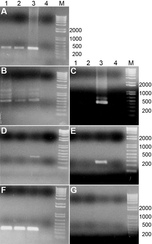
Figure 1. mRNA detection of p53, TAp63, ΔNp63, TAp73, and ΔNp73
Electrophoretic gels showing the presence or absence of mRNA for members of the p53 family of proteins in central and peripheral corneal endothelium. A: p53. B: TAp63. C: ΔNp63. D: TAp73. E: ΔNp73. F: GAPDH (housekeeping protein present in metabolically active cells). G: Samples incubated with water instead of reverse transcriptase (RT) confirmed that DNA contaminants were not present. Lane 1: Central human corneal endothelial cells. Lane 2: Peripheral human corneal endothelial cells. Lane 3: Positive control (human peripheral corneal epithelium and stroma). Lane 4: Negative control (water). Lane M: PCR marker. All bands migrated to their expected product length as summarized in Table 2.