![]() Figure 7 of
Liu, Mol Vis 2005;
11:321-327.
Figure 7 of
Liu, Mol Vis 2005;
11:321-327.
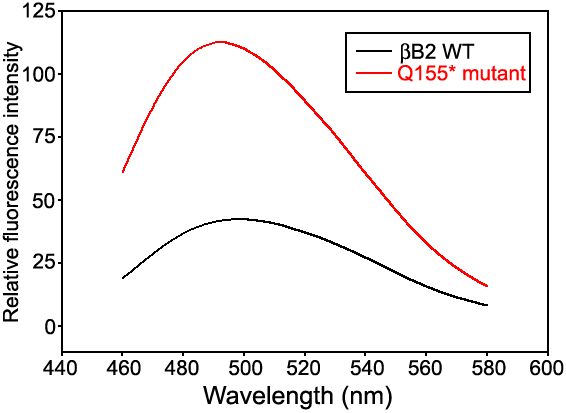
Figure 7. Bis-ANS fluorescence of βB2-crystallin
It can be seen that more hydrophobic surfaces are accessible to the Bis-ANS probe in the B155* mutant than in the WT βB2-crystallin. The excitation wavelength is λex=395 nm. Protein and Bis-ANS concentrations were 0.08 mg/ml and 1x10⁻⁵ M, respectively.