![]() Figure 4 of
Brand, Mol Vis 2005;
11:309-320.
Figure 4 of
Brand, Mol Vis 2005;
11:309-320.
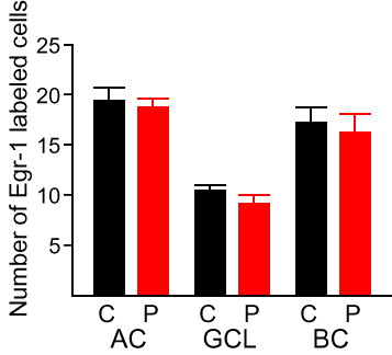
Figure 4. Comparison of Egr-1 protein expression between central and peripheral retina
Density of Egr-1 labeled amacrine cells (AC), ganglion cell layer (GCL), and bipolar cells (BC) in the central (C) and peripheral (P) retina of untreated animals 1 h after light onset in the morning. Cell counts given in the figure correspond to average cell counts in two microscope fields. Error bars represent the standard error of the mean. The sample size is 6 animals per group.