![]() Figure 1 of
Brand, Mol Vis 2005;
11:309-320.
Figure 1 of
Brand, Mol Vis 2005;
11:309-320.
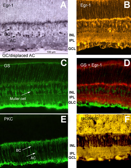
Figure 1. Immunohistochemical labeling of Egr-1, glutamine synthetase (GS), protein kinase C (PKC) and recoverin in the mouse retina
Transverse sections through mouse retina were immunocytochemically labeled. A: Section labeled for Egr-1 using immunoperoxidase coupled diaminobenzidine staining. Cells in the ganglion cell layer (GC), amacrine cells (AC), and putative bipolar cells (BC) in the inner nuclear layer are labeled. B: Section labeled for Egr-1 using immunoperoxidase with a fluorescent second antibody. C: Section immunolabeled for glutamine synthetase (GS). GS labeling is restricted to Müller cells. D: Double labeling for GS and Egr-1 shows that Müller cells are not Egr-1 immunoreactive. E: Section immunolabeled for protein kinase C (PKC). Bipolar cells and some amacrine cells are labeled. The PKC-immunoreactive axons of the bipolar cells terminate in large varicosities in the inner third of the inner plexiform layer (IPL) [32]. F: Section labeled for recoverin. ON and OFF cone bipolar cells are labeled. The inner nuclear layer (INL) and ganglion cell layer (GCL) are also labeled. The scale bar represents 100 μm for all frames.