![]() Figure 7 of
Ash, Mol Vis 2005;
11:298-308.
Figure 7 of
Ash, Mol Vis 2005;
11:298-308.
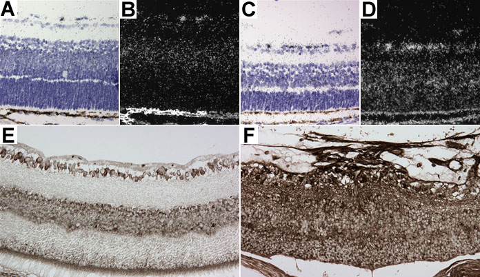
Figure 7. VEGF expression is up regulated in LIF transgenic retinas
VEGF mRNA expression was observed by in situ hybridization (A-D). Brightfield images (A,C) showing the retinal cell layers. Darkfield images (B,D) showing localization of VEGF mRNA expression. In non-transgenic P6 retinas (A,B), the astrocytes and the Müller cells located in the inner nuclear layer were positive. In the LIF transgenic P6 retina (C,D), we observed higher levels of VEGF mRNA expression in Müller cells and astrocytes, but we also observed high expression in photoreceptors and inner retinal neurons. VEGF protein was localized by immunohistochemistry. In normal P14 retinas, VEGF protein was found in the ganglion cell layer (GCL) and in the RPE and choroid (E). In P14 LIF transgenic retinas (F), VEGF protein was markedly increased throughout the retina and in the vascular membrane.