![]() Figure 6 of
Ash, Mol Vis 2005;
11:298-308.
Figure 6 of
Ash, Mol Vis 2005;
11:298-308.
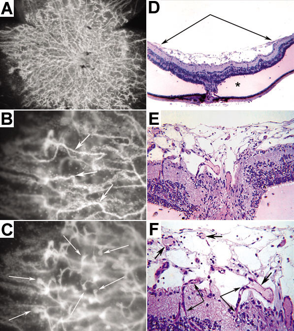
Figure 6. Proliferation of neovascular membranes in older transgenic retinas
An ADPase incubated P21 LIF Tg retina that has been flat embedded and viewed en bloc (A-C). The fellow eye was embedded whole in JB-4 and then sectioned (D-F). A: A dense pre-retinal neovascular membrane. B: Higher magnification of the neovascular membrane in A with focus specifically on the pre-retinal blood vessels (arrows) at the vitreoretinal interface. C: The same area at high magnification but focused on the budding of the vasculature and invasion into the inner retina. D: Retinal detachment due to traction on the retina (double arrows) by the pre-retinal membrane. There is eosinophilic subretinal fluid (*) associated with the retinal detachment. E-F: Higher magnification of a section through the optic nerve showing capillaries invading retina (double arrows) from the pre-retinal neovascular membrane (arrow).