![]() Figure 4 of
Ash, Mol Vis 2005;
11:298-308.
Figure 4 of
Ash, Mol Vis 2005;
11:298-308.
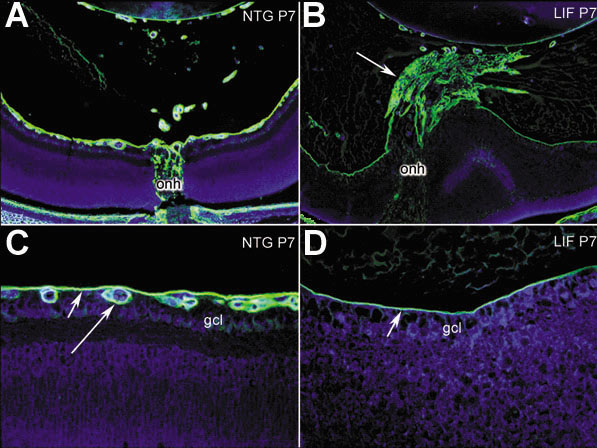
Figure 4. Retinal blood vessels are absent in transgenic mice
P7 retinas were stained by immunofluorescence using an anti-laminin antibody to label the basement membrane of the vasculature and inner limiting membrane of the retina. In non-transgenic eyes (A,C), the blood vessels (C, long arrow) are present in the retina just below the inner limiting membrane (C, short arrow). A tuft of laminin positive tissue was observed near the optic nerve head (onh), which extended into the vitreous to the lens (B, arrow). However, no blood vessels were found in the retina, underneath the inner limiting membrane (D, arrow). "gcl" refers to the ganglion cell layer in C,D.