![]() Figure 2 of
Ash, Mol Vis 2005;
11:298-308.
Figure 2 of
Ash, Mol Vis 2005;
11:298-308.
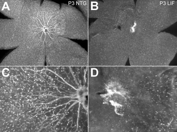
Figure 2. Reduction of ADPase positive cells in transgenic retinas
ADPase incubated retinas from non-transgenic (A,C) and LIF transgenic animals (B,D) at P3. The hyaloid vessels in the vitreous were removed to demonstrate the density of ADPase positive angioblasts and endothelial cells in the retina. Higher magnifications of the optic nerve heads are shown in the lower panels (C,D). We observed marked reduction of ADPase positive cells in the LIF transgenic retinas.