![]() Figure 1 of
Ash, Mol Vis 2005;
11:298-308.
Figure 1 of
Ash, Mol Vis 2005;
11:298-308.
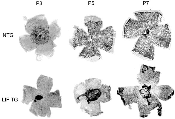
Figure 1. LIF blocks the development of the initial retinal vasculature
Retinas were dissected from the eyes and the blood vessels were stained using Griffonia simplicifolia (GS) lectin. After staining, the retinas were flat mounted on glass slides. The image shows retinas from P3, P5, and P7 mice from non-transgenic retinas (NTG), and LIF transgenic retinas (LIF TG).