![]() Figure 6 of
Rao, Mol Vis 2005;
11:288-297.
Figure 6 of
Rao, Mol Vis 2005;
11:288-297.
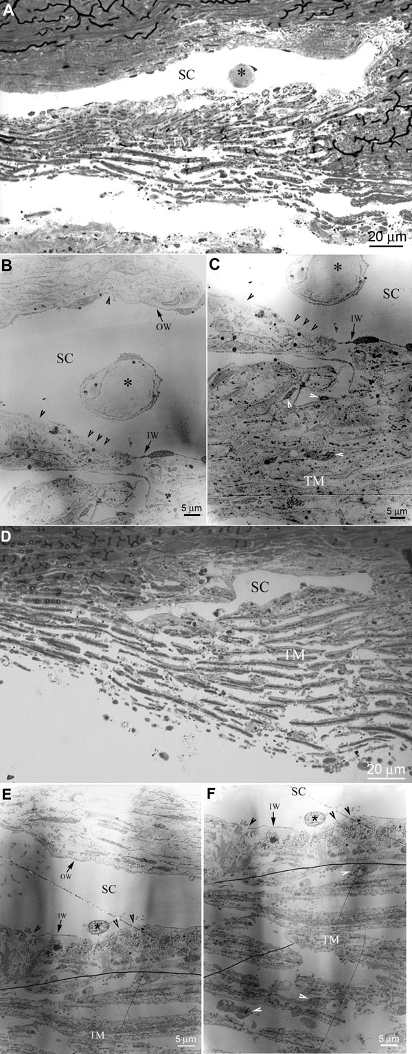
Figure 6. Histological integrity of aqueous humor outflow pathway expressing dominant negative Rho-kinase
A and D illustrate light microscopic morphological changes in the aqueous humor outflow pathway of human eye anterior segments expressing either GFP alone (A) or DNRK and GFP (D). In both specimens analyzed, the morphology of Schlemm's canal (SC) and the trabecular meshwork (TM) appear to be comparable, with no obvious distinction between the control (GFP alone) and experimental (DNRK expressing) specimens. Also, in both specimens we noted a few SC cells that had detached from the subendothelial area of juxtacanicular tissue (indicated with asterisks). However, transmission EM analyses depict obvious discontinuities in the lining of the inner wall (IW) of SC (black arrow heads in B and C) in control (GFP alone) and DNRK expressing specimens (E and F). Both light and EM microscope based analysis revealed some morphological changes such as rounding of TM cells and detachment of TM cells from the TM beams in DNRK infected specimens (indicated with white arrow heads in F). In GFP expressing control specimens, the cells appear long and firmly attached to the TM beams (indicated with white arrow heads in C). The outer wall (OW) of SC is labeled.