![]() Figure 5 of
Rao, Mol Vis 2005;
11:288-297.
Figure 5 of
Rao, Mol Vis 2005;
11:288-297.
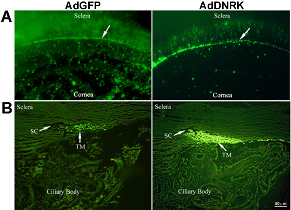
Figure 5. Expression of DNRK in the anterior segment of organ cultured human eyes
Organ cultured anterior segments of human eyes infected with either adenoviral vector (Ad-DNRK-GFP or Ad-GFP), clearly exhibited expression of GFP. A: Expression pattern of GFP in ciliary muscle, anterior chamber angle (arrows), and in corneal endothelium of control (infected with adenoviral vector expressing GFP alone) and experimental (DNRK and GFP) specimens. The expression of GFP was relatively intense in the anterior chamber angle compared to cornea and ciliary muscle in both control and experimental specimens. These data were obtained from perfused anterior segments, at the 6 day after infection time point. B: Expression of GFP in aqueous outflow pathway of control and DNRK samples. Analysis of cryosections derived from anterior segments of eyes infected with Ad-DNRK-GFP or Ad-GFP demonstrate intense expression of GFP in TM tissue relative to ciliary muscle and sclera in both the samples.