![]() Figure 4 of
Rao, Mol Vis 2005;
11:288-297.
Figure 4 of
Rao, Mol Vis 2005;
11:288-297.
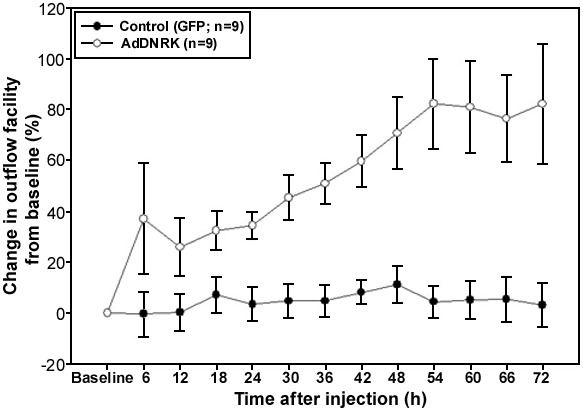
Figure 4. Changes in aqueous humor outflow facility in enucleated human eyes expressing DNRK
Organ cultured anterior segments of human eyes infected with an Ad-DNRK-GFP viral vector (107 pfu) exhibited significantly increased aqueous outflow facility starting at 12 h after infection. At 72 h after infection, the percent change in facility from the base line value was 80% in cells infected with the Ad-DNRK-GFP viral vector (p<0.006). This increase in outflow facility was sustained until 6 days after infection (data not shown). Expressing GFP alone (infected with Ad-GFP viral vector) showed very little change in facility (<5%) from baseline values. A paired two tailed Student's t-test was applied to determine the significance of difference in outflow facility between control and DNRK expressed. Vertical bars represent the standard errors of the mean.