![]() Figure 7 of
Wen, Mol Vis 2005;
11:256-262.
Figure 7 of
Wen, Mol Vis 2005;
11:256-262.
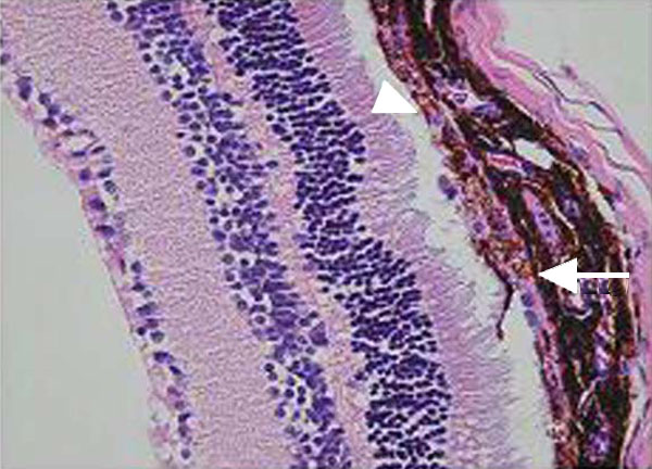
Figure 7. Uptake of donor derived microbeads by recipient RPE
Cultured RPE were incubated with superparamagnetic microbeads and the free beads removed just before the adherent RPE were trypsinized, washed, and passed through a magnet. The RPE, which contained microbeads, were immediately injected into the subretinal of C57BL/6 mice. Three weeks later the eyes were examined by histology. Microbeads are detectable within the RPE of the recipient (arrow) and in the layer formed by the donor RPE (arrow head; 200x).