![]() Figure 4 of
Wen, Mol Vis 2005;
11:256-262.
Figure 4 of
Wen, Mol Vis 2005;
11:256-262.
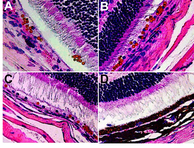
Figure 4. Microbeads in the subretinal space
A bent needle was inserted into a perilimbal incision, angled around the lens, and inserted into the inferior quadrant. The resulting detachment was typically between 6 and 9 o'clock. Each injection consisted of around 105 BSA coated microbeads suspended in 2 μl of HBSS without phenol red. Microbeads in the subretinal space in the Balb/c mouse eye after 1 day (A), 1 week (B), or 3 weeks (C) or in B6 mice after 3 weeks (D). The eyes were examined by histology. The microbeads are clearly seen as symmetrically round, orange objects (400x).