![]() Figure 2 of
Wen, Mol Vis 2005;
11:256-262.
Figure 2 of
Wen, Mol Vis 2005;
11:256-262.
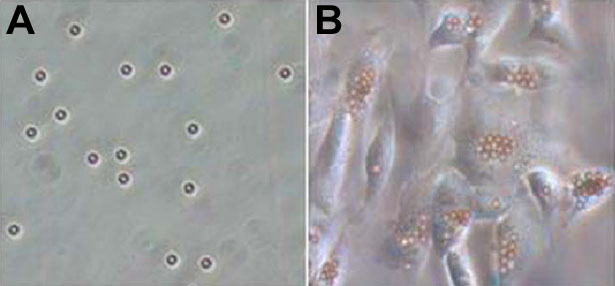
Figure 2. Microbeads in vitro
Microbeads were incubated with adherent cultures of RPE and nonadherent microbeads removed by washing the monolayer. Microbeads (A) and RPE with ingested microbeads (B) were photographed under phase contrast microscopy (200x).