![]() Figure 1 of
Wen, Mol Vis 2005;
11:256-262.
Figure 1 of
Wen, Mol Vis 2005;
11:256-262.
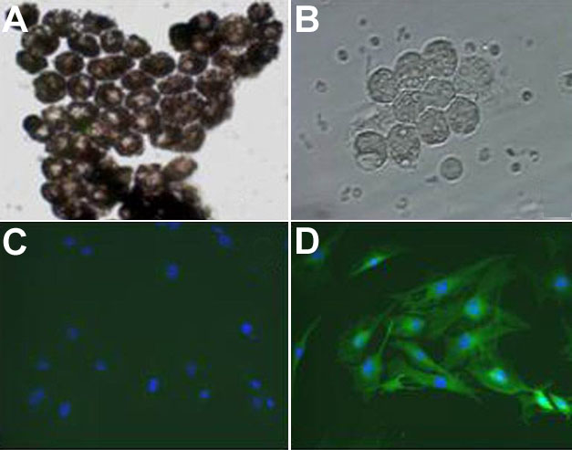
Figure 1. Characterization of mouse RPE
RPE were isolated from mouse eyes by digestion with proteolytic enzymes. Pigmented C57BL/6 RPE are shown by light microscopy (A) whereas nonpigmented Balb/c RPE are shown by phase contrast microscopy (B). Freshly isolated RPE displayed typical hexagonal morphology and contained oil droplets. After several weeks in culture, RPE were incubated with an isotype control antibody (C) or an anticytokeratin antibody (Abcam) that reacts with cytokeratin from a variety of species from lizards to mice and humans (D) followed by fluorescent anti-IgG and DAPI, to stain the nuclei. Virtually all cells reacted with the anti-cytokeratin antibody.