![]() Figure 5 of
Wu, Mol Vis 2005;
11:28-35.
Figure 5 of
Wu, Mol Vis 2005;
11:28-35.
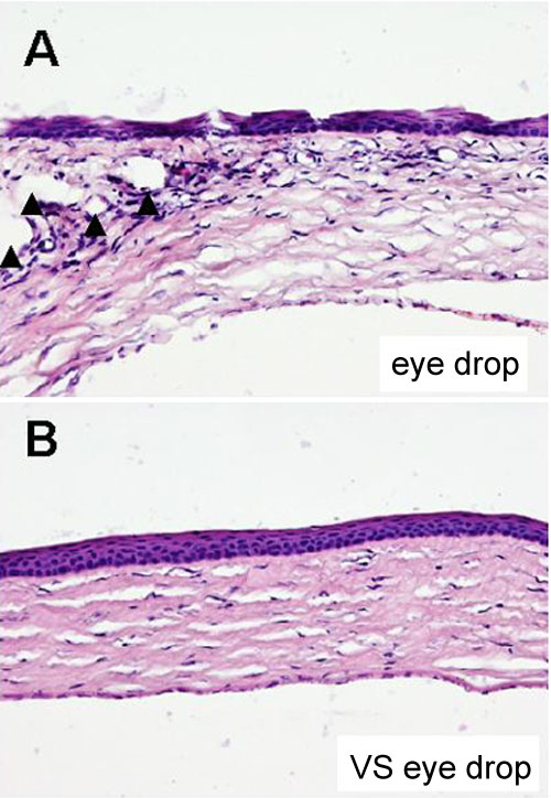
Figure 5. Histological analysis of basic fibroblast growth factor (bFGF) implanted rat cornea after eye drop treatment
After eye drop treatment for 7 days, the bFGF implanted rat corneas were harvested and stained with hematoxylin and eosin (200x). In eyes that received the control eye drop (A), multiple lumen-like formations containing red blood cells presented in the corneal stroma (arrowhead) with the bFGF implant. Neocapillaries (arrowheads) are prominent in the corneal stroma. The intervening stroma exhibits edema and a mononuclear inflammatory response. There is no significant neovascularization shown in eyes receiving VS eye drops (B).