![]() Figure 7 of
Beltran, Mol Vis 2005;
11:232-244.
Figure 7 of
Beltran, Mol Vis 2005;
11:232-244.
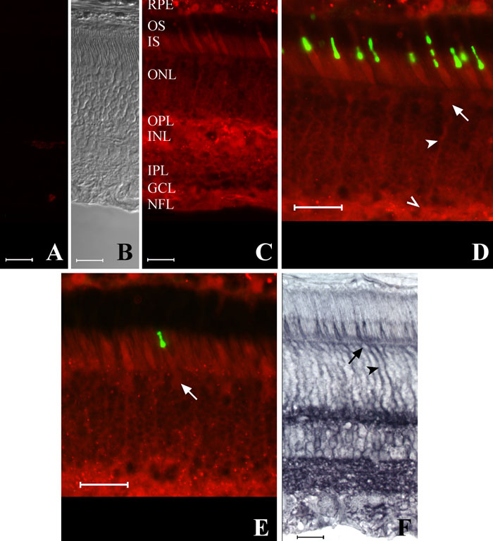
Figure 7. Immunolocalization of CNTFRα in cone photoreceptor cells of the dog
A: Negative control. B: Negative control with DIC optics. C: Immunofluorescence labeling with the anti-chick CNTFRα antibody. D,E: Double immunofluorescence labeling (overlaid images) with the anti-chick CNTFRα (red) and COS1 (green, D) or OS2 (green, E) antibodies. F: Pattern of immunoenzymatic labeling with the anti-human CNTFRα antibody. Intense labeling was present at the level of the inner segments (IS) of both rod and cones, outer plexiform layer (OPL), inner nuclear layer (INL), ganglion cell layer (GCL), and nerve fiber layer (NFL). CNTFRα immunolabeling of M/L (D) and S (E) cones was localized to their inner segments, cell bodies (arrows), axons (arrowheads), and pedicles (open arrowhead). Scale bars represent 20 μm.