![]() Figure 6 of
Beltran, Mol Vis 2005;
11:232-244.
Figure 6 of
Beltran, Mol Vis 2005;
11:232-244.
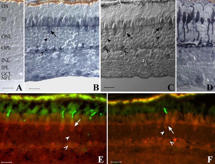
Figure 6. Immunolocalization of CNTFRα in the adult human retina
A: Negative control (pigmented RPE). B: Pattern of immunoenzymatic labeling with the anti-chick CNTFRα antibody. C: Pattern of immunoenzymatic labeling with the anti-rat CNTFRα antibody. D: Sequential section (to B) labeled with human cone arrestin antibody. E,F: Double immunofluorescence labeling (overlaid images) with the anti-chick CNTFRα (red) and COS1 (green, E) or OS2 (green, F) antibodies. Intense labeling with the CNTFRα antibody (B) was seen at the inner segments (IS), outer nuclear layer (ONL; cone cell bodies), outer plexiform layer (OPL), ganglion cell layer (GCL), and nerve fiber layer (NFL). Labeling was present (B,C,E,F) at rod and cone IS, cone cell bodies (arrows), axons (arrowheads), and pedicles (open arrowheads). Both M/L (E) and S (F) cones were labeled by the anti-chick CNTFRα antibody. Scale bars represent 20 μm.