![]() Figure 5 of
Beltran, Mol Vis 2005;
11:232-244.
Figure 5 of
Beltran, Mol Vis 2005;
11:232-244.
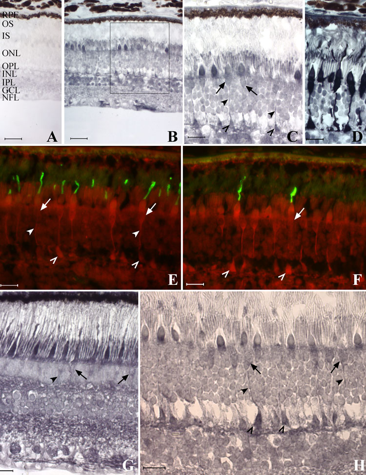
Figure 5. Immunolocalization of CNTFRα in the adult monkey retina
A: Negative control (pigmented RPE). B: Pattern of immunoenzymatic labeling in the rhesus macaque with the anti-chick CNTFRα antibody. C: Boxed region in B. D: Sequential section labeled with human cone arrestin antibody. E,F: Double immunofluorescence labeling (overlaid images) in the rhesus macaque with the anti-chick CNTFRα (red) and COS1 (green, E) or OS2 (green, F) antibodies. G: Pattern of immunoenzymatic labeling in the cynomolgus macaque with the monoclonal anti-human CNTFRα antibody. H: Pattern of immunoenzymatic labeling in the rhesus macaque with the anti-rat CNTFRα antibody. Intense labeling with the CNTFRα antibodies was seen at the inner segments (IS), outer nuclear layer (ONL; cone cell bodies), outer plexiform layer (OPL), inner nuclear layer (INL), ganglion cell layer (GCL), and nerve fiber layer (NFL; B,G). Labeling was present at both rod and cone IS, and at cone cell bodies (arrows), axons (arrowheads), and pedicles (open arrowheads; C,E-H). Both M/L (E) and S (F) cones were labeled by the CNTFRα antibody. Scale bars represent 40 μm (A,B) or 20 μm (C-H).