![]() Figure 3 of
Beltran, Mol Vis 2005;
11:232-244.
Figure 3 of
Beltran, Mol Vis 2005;
11:232-244.
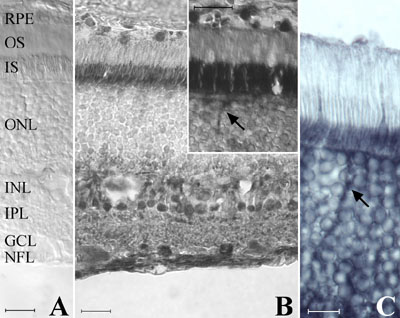
Figure 3. Immunolocalization of CNTFRα in the adult cat retina
A: Negative control. B: Pattern of immunoenzymatic labeling with the anti-chick CNTFRα antibody. C: Immunoenzymatic labeling of photoreceptors with the anti-human CNTFRα antibody. Intense labeling with the anti-chick CNTFRα antibody was seen at the retinal pigment epithelium (RPE), inner segments (IS), inner nuclear layer (INL), ganglion cell layer (GCL), and nerve fiber layer (NFL; B). With longer incubation times in the enzyme substrate (DAB), labeling of cone cell bodies and their extending axons (arrow) could be detected (inset to B, C). Scale bars represent 20 μm (A,B) or 10 μm (C).