![]() Figure 1 of
Beltran, Mol Vis 2005;
11:232-244.
Figure 1 of
Beltran, Mol Vis 2005;
11:232-244.
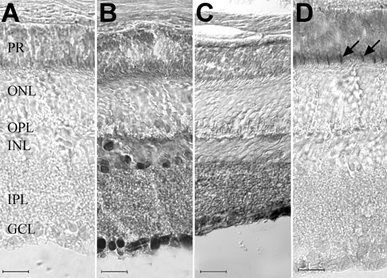
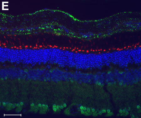
Figure 1. Immunolocalization of CNTFRα in the adult mouse retina
A: Negative control. B: Pattern of immunoenzymatic labeling with the anti-chick CNTFRα antibody. C: Pattern of immunoenzymatic labeling with the anti-rat CNTFRα antibody. D: Sequential section (to B) labeled with mouse cone arrestin antibody. E: Immunofluorescence labeling (overlaid images) with the anti-chick CNTFRα antibody (green), DAPI (blue), and peanut agglutinin (red). Intense labeling with the CNTFRα antibodies (B,E) was limited to ganglion cells, nerve fibers, and cells located predominantly at the vitreal and scleral borders of the inner nuclear layer (INL). Less intense labeling was also present at the inner plexiform layer (IPL) and outer plexiform layer (OPL). Nonspecific staining was observed at the photoreceptor layer (A-C) and was distinct from the specific cone inner segment labeling (arrows in D) obtained with the mouse cone arrestin antibody. Fluorescence immunocytochemistry confirmed the absence of photoreceptor labeling with the anti-chick CNTFRα antibody (E). Scale bars represent 20 μm (A-D) or 40 μm (E).