![]() Figure 2 of
Garcia-Valenzuela, Mol Vis 2005;
11:225-231.
Figure 2 of
Garcia-Valenzuela, Mol Vis 2005;
11:225-231.
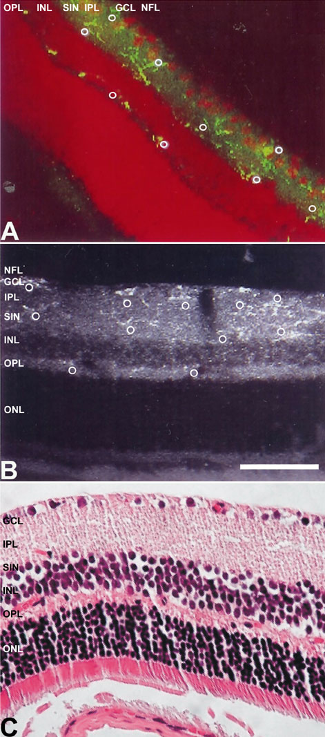
Figure 2. Microglia in various depths of retinal cross-sections after axotomy
A: Confocal photographs of rat retina at 18 days after axotomy, reacted with OX42 antibody (green) showing microglia (white "o") in and between the ganglion cell layer (GCL), superficial inner nuclear layer (SINL), and outer plexiform layer (OPL) in transverse sections of rat retina. The retina was counterstained with propidium iodide (red). B: Confocal photographs of rat retina at 18 days after axotomy, reacted with OX42 antibody (white) showing microglia (white "o"). C: A section of normal retina stained with hematoxilin-eosin is shown for histological reference. Scale bar represents 180 μm.