![]() Figure 1 of
Garcia-Valenzuela, Mol Vis 2005;
11:225-231.
Figure 1 of
Garcia-Valenzuela, Mol Vis 2005;
11:225-231.
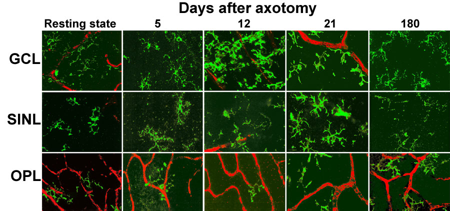
Figure 1. Progressive changes of microglia induced by axotomy in the three layers of retinal flatmounts
Confocal pictures from rat retinas reacted with OX42 antibody, showing microglia (green) in the GCL (top row), SINL (middle row), and OPL (bottom row). Notice their progressive transformation from resting state, 5 days after axotomy, to their peak of activity at 12 days after axotomy, and subsequent return towards a resting state at 21 days after axotomy and 180 days after axotomy. Blood vessels appear colorized in red. Scale bar represents 50 μm.