![]() Figure 3 of
Rollin, Mol Vis 2005;
11:216-224.
Figure 3 of
Rollin, Mol Vis 2005;
11:216-224.
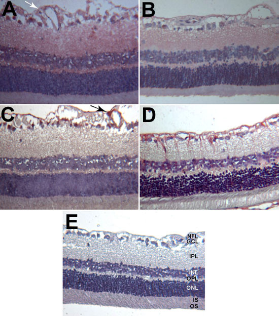
Figure 3. ANP and GFAP immunoreactivities in the control and diabetic rat retinas
Photomicrographs of control retina showing ANP (A) and GFAP (C) immunostaining and of diabetic retina after 3 months of diabetes showing ANP (B) and GFAP (D) immunostaining. ANP and GFAP immunoreactivities could be observed on astrocytes and their processes around blood vessels in adjacent sections of the retina (A,C, arrows). Negative controls for the immunohistochemical detection of ANP (E) were free of labeling. All the sections were counterstained with haematoxylin and photographed at a magnification of 160x. The photoreceptor outer segments (OS), photoreceptor inner segments (IS), outer nuclear layer (ONL), outer plexiform layer (OPL), inner nuclear layer (INL), inner plexiform layer (IPL), ganglion cell layer (GCL), and nerve fiber layer (NFL) are labeled in E.