![]() Figure 3 of
Kerrison, Mol Vis 2005;
11:208-215.
Figure 3 of
Kerrison, Mol Vis 2005;
11:208-215.
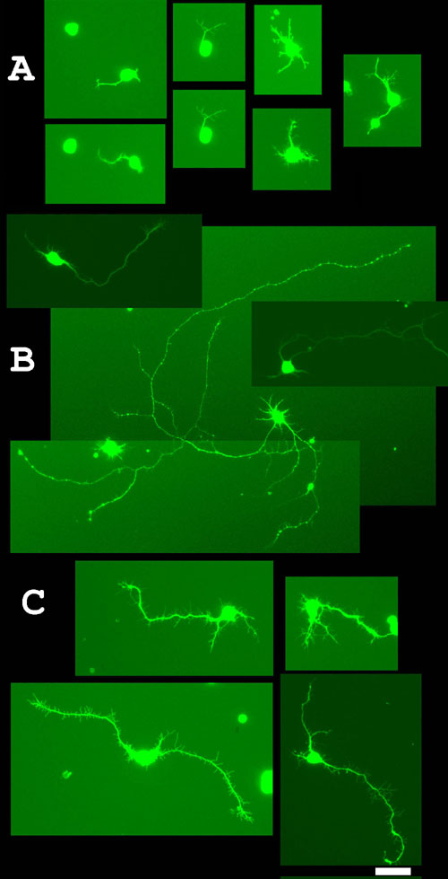
Figure 3. Neurite outgrowth of cultured RGCs
At 120 h, neurites were visualized after staining with Calcein AM. RGCs were grown in the standard media alone (A), in the presence of BDNF (B), or in the presence of BMP2 (C). In the presence of BDNF, 15 to 30% of cells were "outgrowth neurons," having neurite features surpassing a threshold of one standard deviation above the mean neurite characteristic of RGCs cultured in growth medium alone. The scale bar represents 40 μm.