![]() Figure 2 of
Kerrison, Mol Vis 2005;
11:208-215.
Figure 2 of
Kerrison, Mol Vis 2005;
11:208-215.
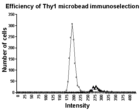
Figure 2. Efficient Thy-1 immunoselection
Both the cells passing through the magnetized columns (represented as open squares in the figure) and the cells retained by the magnetized column and later eluted (represented as filled triangles in the figure) were plated, immunolabeled with Thy-1 antibody and secondary FITC conjugated antibody, and imaged with the Cellomics KSR instrument. The intensity of fluorescence for each cell is plotted, demonstrating two distinct populations of cells (p<0.00001, Student's t-test) and highly efficient immunomagnetic purification. The fluorescence intensity of the cells passing through the magnetized columns (represented as open squares in the figure) can be considered background.