![]() Figure 2 of
Hayashi, Mol Vis 2005;
11:201-207.
Figure 2 of
Hayashi, Mol Vis 2005;
11:201-207.
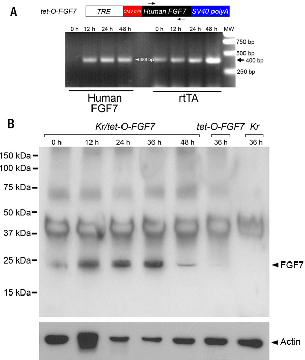
Figure 2. Induction of FGF7 transgene expression
A: Diagram of tet-O-FGF7 minigene and RT-PCR products of hFGF7 and rtTA. Arrows in the diagram indicate the position of primers used in RT-PCR for hFGF7. The cDNA prepared by RT from corneas of bitransgenic mice at 0, 12, 24, and 48 h after an intraperitoneal injection of doxycycline were examined. mRNA of rtTA was detected with or without doxycycline treatment, while the mRNA of FGF7 was only detected upon induction for 12-48 h. TRE, tetracycline responsive element; CMVmin, cyotmegarovirus minimum promoter. B: Western blotting analysis of the expression of FGF7 and actin. Corneas excised from Kr/tet-O-FGF7 bitransgenic, and Kr and/or tet-O-FGF7 single transgenic mice are examined. Peak expression of FGF7 was observed in bitransgenic mice at 36 h after an intraperitoneal injection of doxycycline. FGF7 was also detected at lower levels in bitransgenic mice at 12, 24, and 48 h after doxycycline admindstration. A small amount of FGF7 protein was also detected in single transgenic mice at 36 h of doxycycline induction.