![]() Figure 1 of
Hayashi, Mol Vis 2005;
11:201-207.
Figure 1 of
Hayashi, Mol Vis 2005;
11:201-207.
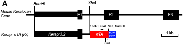
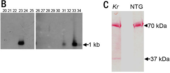
Figure 1. Preparation of kerapr-rtTA transgenic mice
A: Diagram of the endogenous mouse keratocan gene and the Kerapr-rtTA minigene. Arrows indicate the primers used for RT-PCR to detect transgene expression. A 3.2 kb fragment of the first exon and 5' promoter region of the keratocan gene was used to direct rtTA expression. This minigene was used to generate Kerapr-rtTA transgenic mice using standard transgenesis techniques [12]. B: Southern blot hybridization identified four transgenic founder lines (Kr23, Kr31, Kr33, Kr34). C: Western blotting analysis of the expression of rtTA. NTG indicates the non-transgenic littermate. The anti-VP-16 antibody detected rtTA (37 kDa, arrow) and also an unknown cellular protein with a molecular weight at 70 kDa (arrowhead) as described by the manufacturer. Only line Kr23 showed positive rtTA protein expression.