![]() Figure 4 of
Yamagami, Mol Vis 2005;
11:192-200.
Figure 4 of
Yamagami, Mol Vis 2005;
11:192-200.
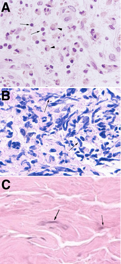
Figure 4. Hematoxylin-eosin stainings in giant papillae of representative AKC patient (patient 1) and patient 5
A: Hematoxylin-eosin staining shows that infiltration of mononuclear cells (arrows) and eosinophils (arrowheads) are predominant. B: Marked fibroblast-like cell (arrows) rather than mononuclear and eosinophil infiltrations are observed in giant papillae of patient 5. C: Some fibroblast-like cells (arrows) are present under normal conjunctival epithelium. Original magnification was 200x.