![]() Figure 2 of
Yamagami, Mol Vis 2005;
11:192-200.
Figure 2 of
Yamagami, Mol Vis 2005;
11:192-200.
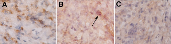
Figure 2. Immunohistochemical expression of TARC/CCL17 and MDC/CCL22 in the giant papillae of an AKC patient
TARC/CCL17 and MDC/CCL22 were expressed in the giant papillae of all three available patients examined for immunohistochemistry (patients 1, 2, and 4). A: A representative photograph is shown (patient 2). TARC/CCL17 positive staining is present in the subepithelial part of the conjunctiva. B: MDC/CCL22 positive cells in the conjunctivae were detected, but were rare in each giant papilla of the 3 patients (Arrow). C: No positive staining is detected in the AKC conjunctivae with non-immunized control IgG. Positive staining is not detected in the normal conjunctivae (data not shown). Original magnification was 200x.