![]() Figure 6 of
Normand, Mol Vis 2005;
11:184-191.
Figure 6 of
Normand, Mol Vis 2005;
11:184-191.
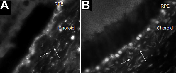
Figure 6. In vivo light controlled distribution of ODNs in the choroid after intravitreal injection of vectosomes in the rat eye
Fluorescence photomicrographs of sections showing the choroid cells 24 h after intravitreal injections of vectosomes. A: Vectosomes were dispersed in the choroid cells as punctate fluorescent staining (white arrows) prior to light treatment. B: After illumination, nuclear fluorescence was observed in the choroid cell nuclei (white arrows). An anti c-raf kinase ODN was used. The ODN was HEX labeled.