![]() Figure 3 of
Normand, Mol Vis 2005;
11:184-191.
Figure 3 of
Normand, Mol Vis 2005;
11:184-191.
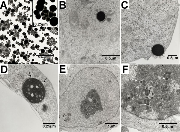
Figure 3. Electron photomicrograph of VP22/F-ODN complexes in suspension and OCM-1 cells incubated with the complexes
VP22/F-ODN complexes were applied to EM grids covered by formvar support films and examined by electron microscopy. A: Regular spherical particles could be observed. B: VP22/ODN complexes could be observed as electron dense material in the cell cytoplasm. C: The initial step of cell internalization could be recorded with the cell membrane forming invaginations around the electron dense particles. D: Illumination affected the particles leading to disruption of the membrane around the particles. A lace-like structure became visible within the particles. E,F: Along with these changes, spotty materials could be detected only inside nuclei of cells treated with light. These materials might come from the disruption of the vectosomes, enabling accumulation of smaller fragments in the cell nuclei.