![]() Figure 2 of
Normand, Mol Vis 2005;
11:184-191.
Figure 2 of
Normand, Mol Vis 2005;
11:184-191.
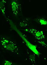
Figure 2. Intracellular redistribution of F-ODN following illumination of OCM-1 cells loaded with vectosomes
OCM-1 cells were incubated overnight at 37 °C with VP22/F-ODN complexes as described under Methods. The cells were washed in fresh medium and illuminated 10 s with the Hg lamp. A time series was recorded every 10 s by confocal microscopy over 2 min and is presented as a movie.
Note that the slide bar at the bottom of the quicktime movie can be used to manually control the flow of the movie. If you are unable to view the movie, a representative frame is included below.
| This animation requires Quicktime 6 or later. Quicktime is available as a free download. |