![]() Figure 1 of
Normand, Mol Vis 2005;
11:184-191.
Figure 1 of
Normand, Mol Vis 2005;
11:184-191.
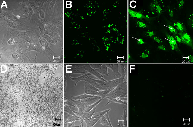
Figure 1. VP22 mediated delivery of F-ODN into OCM-1 cells: uptake and light induced redistribution
OCM-1 cells were incubated overnight at 37 °C with either VP22/F-ODN complexes (A-D) or with free F-ODN (E,F). Images of living cells were captured by confocal microscopy immediately following (A,B) and 10 s after (C) illumination with the Hg lamp. The illumination triggered ODN redistribution within the cells and images were captured 2 min later. After illumination the fluorescent particles were disrupted leading to a diffuse staining in the cytoplasm and the nuclei (C). Cells loaded with VP22/F-ODN complexes were recorded by phase microscopy (A,D) and presented their typical elongated morphology. Magnifications is x63 in A,B,C,E,F and x5 in D.