![]() Figure 9 of
Montiani-Ferreira, Mol Vis 2005;
11:11-27.
Figure 9 of
Montiani-Ferreira, Mol Vis 2005;
11:11-27.
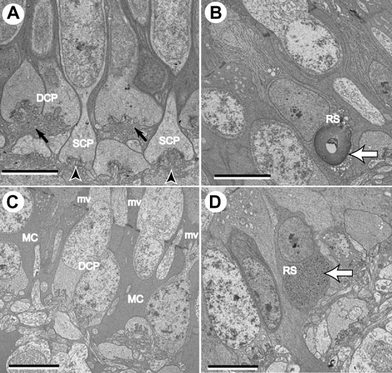
Figure 9. Abnormalities of the outer nuclear layer
Ultra-thin sections of retinal samples from rge/rge and control birds at 60 days of age. A: Note the aspect of the normal chick OPL at 60 days of age. The double cone pedicles terminate in the outer sublayer of the OPL (small black arrows), whereas the single cone pedicles terminate in the inner sublayer (black arrowheads). B: Occasionally whorls of membranes forming densely packed stacks of coaxial cylindrical bilayers (myelinoid figures, white arrow) were observed in rod cell bodies in the ONL of rge/rge birds. C: Müller cell processes (MC) occupy gaps between photoreceptors cell bodies. Abundant long Müller cells microvilli (mv) are present between the ISs of the photoreceptor cells. D: One large glycogen deposit can be observed in an abnormal location (white arrow). Normally, glycogen can only be found in the ISs. Each bar represent 5 μm. The single cone pedicle (SCP), double cone pedicle (DCP), and rod spherule (RS) are labeled.