![]() Figure 8 of
Montiani-Ferreira, Mol Vis 2005;
11:11-27.
Figure 8 of
Montiani-Ferreira, Mol Vis 2005;
11:11-27.
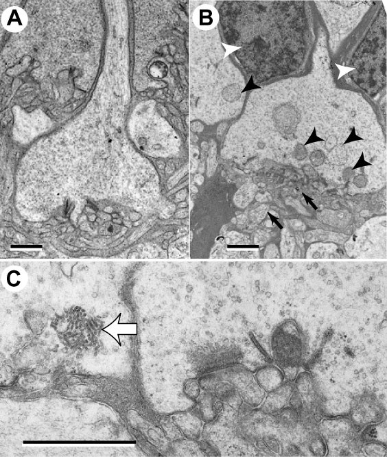
Figure 8. Abnormalities of synaptic terminals
A Synaptic terminal of a cone pedicle of a control chick at 7 days of age. B: A typical example of a synaptic terminal of a cone pedicle of an rge/rge chick at 7 days of age. The cytoplasm is less densely stained. Note the disruption in the architecture of the synaptic terminals (small black arrows). Also note the presence of electron dense glial cell bodies separating the photoreceptors (white arrowheads) and multivesicular bodies (black arrowheads). C: Higher power detail of an rge/rge retinal section at 7 days of age demonstrating one of the sets of numerous flattened (tubuliform) vesicles (wide white arrow) in a cone pedicle. Each bar represents 1 μm.