![]() Figure 7 of
Montiani-Ferreira, Mol Vis 2005;
11:11-27.
Figure 7 of
Montiani-Ferreira, Mol Vis 2005;
11:11-27.
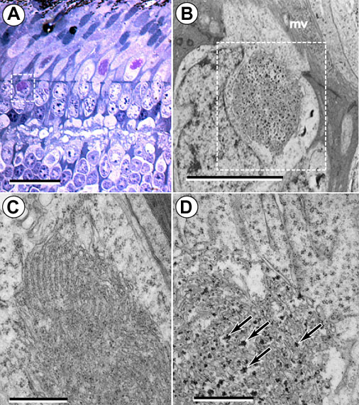
Figure 7. Mislocalization of glycogen deposits
Semi-thin (A) and ultra-thin (B,C,D) sections of rge/rge (A,B,D) and control (C) retina at 7 days of age. A: Semi-thin section showing the detail of a large glycogen deposit (dashed white square) in the perinuclear cytoplasm. Note the disrupted OPL. The bar represents 20 μm. B: Ultrastructural detail of a deposit in the perinuclear cytoplasm of an accessory cell of double cone of an rge/rge bird where it is possible to observe the abnormal accumulation of glycogen (dashed white square). The bar represents 5 μm. An electron dense glial process of a Müller cell (mv) located between the ISs of the photoreceptor cells is indicated. C: Control retina showing evenly arranged SER in the IS (paraboloid). Small glycogen granules can be seen among the cisterns. The bar represents 1 μm. D: Higher magnification of section in B to show abnormal accumulations of glycogen associated with ER (black arrows). The bar represents 1 μm.