![]() Figure 5 of
Montiani-Ferreira, Mol Vis 2005;
11:11-27.
Figure 5 of
Montiani-Ferreira, Mol Vis 2005;
11:11-27.
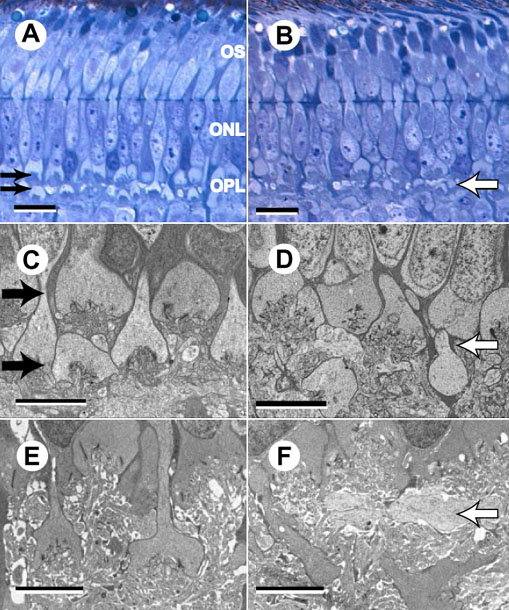
Figure 5. Morphological abnormalities of the outer plexiform layer
Semi-thin (A,B; toluidine blue) and ultra-thin (C,D) retinal sections of a control (A,C) and of an rge/rge chick (B,D), at 1 day of age. E,F are ultra-thin sections of a control (E) and an rge/rge chick (F), at 60 days of age. A: Semi-thin section of a control chick retina demonstrating the detail of the well organized outer OPL. The normal 2 layer arrangement of the photoreceptor synaptic terminals is indicated (two black arrows). B: Note the dilated photoreceptor cell bodies and the disorganization of the OPL architecture in the sample from an rge/rge chick (white arrow). C: The typical 2 layer arrangement of the photoreceptor synaptic terminals is clearly shown here (two black arrows). D: The two layer arrangement of the photoreceptor terminals is lost (white arrow); they were seen in C (two black arrows). Note the disruption of the photoreceptor pedicles and spherules. E,F: The two layer arrangement of the photoreceptor pedicles and spherules is even further disorganized (white arrow) in the rge/rge section compared to controls at 60 days of age. Note that there are fewer synaptic ribbons in the rge/rge section (F) compared to control (E) at this age. In A,B, the bars represent 10 μm. In C-F, the bars represent 5 μm. The outer segments (OS), outer nuclear layer (ONL), and outer plexiform layer (OPL) are labeled.