![]() Figure 3 of
Montiani-Ferreira, Mol Vis 2005;
11:11-27.
Figure 3 of
Montiani-Ferreira, Mol Vis 2005;
11:11-27.
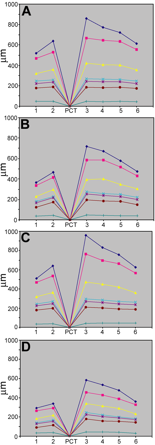
Figure 3. Regional retinal thickness comparison
Mean retinal thickness of each of the different retinal layers from rge/rge (B,D) and control (A,C) birds at two representative age groups. By 56 days of age (A,B; 6 rge/rge, 4 controls), mean thickness of NFL plus GCL (blue), IPL (pink), INL (yellow) and also OS plus IS (brown) layer were already decreased. Note that the ventral regions (1 and 2) and the far dorsal peripheral region (6) thinned the most. In retinas from birds at 270 days of age (C,D; 4 rge/rge, 5 controls), the thickness of the NFL plus GCL, IPL and IS plus OS retinal layers was markedly decreased. The other colored lines represent the retinal pigment epithelium (aquamarine), outer nuclear layer (purple), outer plexiform layer (turquoise), and pecten (PCT).