![]() Figure 16 of
Montiani-Ferreira, Mol Vis 2005;
11:11-27.
Figure 16 of
Montiani-Ferreira, Mol Vis 2005;
11:11-27.
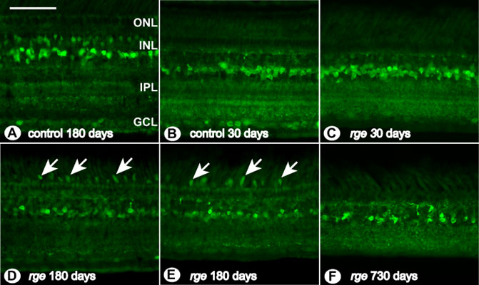
Figure 16. Abnormalities in glycine immunohistochemistry
Glycine immunohistochemistry of sections from retinal samples. A: Control bird at 180 days of age. B: control chick at 30 days of age. C: rge/rge bird at 30 days of age. D: rge/rge chick at 180 days of age (central area of the retina). E: Retinal section from an rge/rge bird at 180 days of age (periphery of the retina). F: Retinal section of an rge/rge bird at 730 days of age. Note the glycine immunoreactive "blobs" (arrows) detected in the ISs of some photoreceptors in retinas from older rge/rge birds (D,E). The bar represents 50 μm. The outer nuclear layer (ONL), inner nuclear layer (INL), inner plexiform layer (IPL), and ganglion cell layer (GCL) are labeled.