![]() Figure 15 of
Montiani-Ferreira, Mol Vis 2005;
11:11-27.
Figure 15 of
Montiani-Ferreira, Mol Vis 2005;
11:11-27.
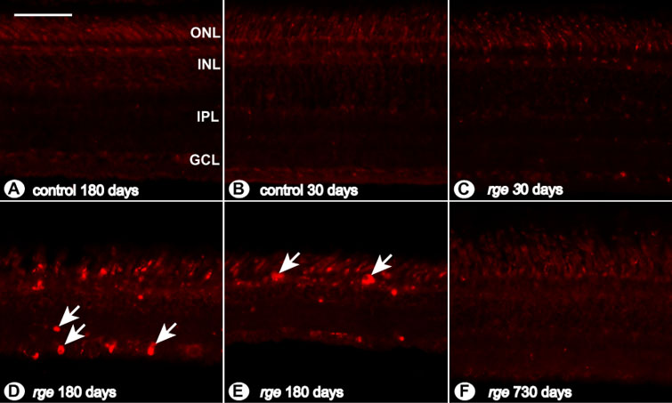
Figure 15. Abnormalities of LEP-100 immunohistochemistry
LEP-100 immunohistochemistry staining of sections from retinal samples. A: Retinal section from a control bird at 180 days of age. B: Control chick at 30 days of age. C: rge/rge chick at 30 days of age. D: Mid-peripheral retinal section of an rge/rge bird at 180 days of age. E: Far-peripheral retina of an rge/rge bird at 180 days of age. F: Central retinal section of an rge/rge bird at 730 days of age. Note the difference between rge/rge (C,D,E) and control (A,B) samples. Several layers of the rge/rge retinas, mainly outer nuclear layer (ONL), inner nuclear layer (INL), inner plexiform layer (IPL), and ganglion cell layer (GCL) at 180 days of age were positively stained for this antibody (white arrows). At 30 days and 730 days of age, no differences could be detected between rge/rge and control samples. The bar represents 50 μm.