![]() Figure 14 of
Montiani-Ferreira, Mol Vis 2005;
11:11-27.
Figure 14 of
Montiani-Ferreira, Mol Vis 2005;
11:11-27.
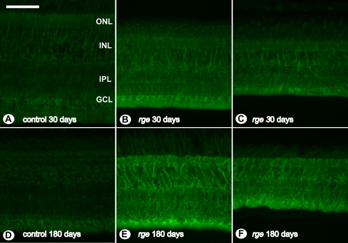
Figure 14. Abnormalities in GFAP immunohistochemistry
GFAP immunohistochemistry staining of sections from retinal samples. A: Control bird at 30 days of age. B: rge/rge chick at 30 days of age. C: Peripheral retina of an rge/rge chick at 30 days of age. D: Control bird at 180 days of age. E: rge/rge bird at 180 days of age. F: Peripheral retina of an rge/rge bird at 180 days of age. Note the rge/rge retinas appear to have a progressive increase of GFAP expression, indicating that glial cells (Müller cells) are reactive. The bar represents 50 μm. The outer nuclear layer (ONL), inner nuclear layer (INL), inner plexiform layer (IPL), and ganglion cell layer (GCL) are labeled.