![]() Figure 13 of
Montiani-Ferreira, Mol Vis 2005;
11:11-27.
Figure 13 of
Montiani-Ferreira, Mol Vis 2005;
11:11-27.
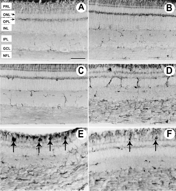
Figure 13. Abnormalities in RCA-1 immunohistochemistry
Immunohistochemically stained retinal sections using RCA-1 antibody of control (A) and rge/rge chicks at 13 days (B), 33 days (C), and 92 days (D-F) of age. A: Most ramified microglia are located in the nerve fiber layer (NFL), inner plexiform layer (IPL) and outer plexiform layer (OPL). There was no significant difference in microglial morphology between control and rge/rge retinas at 13 days of age. C: In rge/rge retinas from 33 day old chicks, some microglia in IPL and OPL showed amoeboid shape indicating activation of these cells. (D-F) Retinal samples from rge/rge birds at 92 days of age. Note many amoeboid microglia with stout processes are seen in INL and NFL (D) and RCA-1 labeled cells similar to macrophages (E) or microglia (F) were present in the photoreceptor layer (black arrows). The bar represents 50 μm. The photoreceptor layer (PRL), outer nuclear layer (ONL), inner nuclear layer (INL), and ganglion cell layer (GCL) are labeled.