![]() Figure 12 of
Montiani-Ferreira, Mol Vis 2005;
11:11-27.
Figure 12 of
Montiani-Ferreira, Mol Vis 2005;
11:11-27.
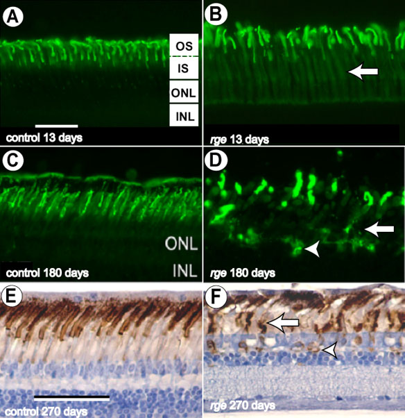
Figure 12. Abnormalities in opsin immunohistochemistry
Fluorescence photomicrographs (A-D) and conventional photomicrograph using brown chromogen (E,F) from immunohistochemistry sections using mouse anti-rhodopsin (A-D) and monoclonal antibody against mouse opsin (E,F). Sections from the mid-periphery of the retina from a control (A) and an rge/rge bird (B), at 13 days of age; from a control (C) and rge/rge bird (D), at 180 days of age, and from a control (E) and rge/rge bird (F), at 270 days of age. Note the presence of increasing amounts of rhodopsin immunoreactivity in the inner segments (IS) of the rge/rge samples (white arrows on B,D,F). Note that the opsin labeling in the ISs of control samples is minimal (A,C,E). Also, note that, compared to controls (C,E), the rod outer segments (OS) in the rge/rge retinas of older birds (D,F) appear swollen, with loss of the normal architecture and organization. The OSs of rge/rge chicks also appeared more widely spaced in comparison to the controls. Mislocalization of opsin is present in rge/rge retinal samples at the outer nuclear layer (ONL)/outer plexiform layer (OPL) level (white arrowheads on D,F). The inner nuclear layer (INL) is also labeled. The bars represents 50 μm.