![]() Figure 11 of
Montiani-Ferreira, Mol Vis 2005;
11:11-27.
Figure 11 of
Montiani-Ferreira, Mol Vis 2005;
11:11-27.
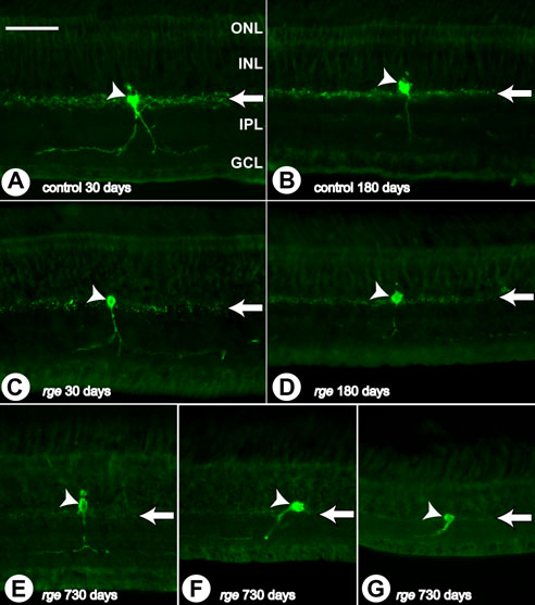
Figure 11. Abnormalities in tyrosine hydroxylase immunohistochemistry
Immunohistochemistry sections using tyrosine hydroxylase (TH) antibody performed on samples from rge/rge and control birds at 30, 180, and 730 days of age. A: Control bird at 30 days of age. B: Control bird at 180 days of age. C: rge/rge bird at 30 days of age. D: rge/rge bird at 180 days of age. E: Central retina of rge/rge bird at 730 days of age. F: Peripheral retina. G: Far peripheral retina. Note that in control retinas, TH positive neurites are found concentrated at the outer border of the IPL, close to the corresponding amacrine cell body. Further neurites from these cells can be observed, at a lesser density, branching bilaterally and extending into the IPL close to the GCL. At later stages of development (180 and 730 days of age) there is an obvious decrease in the density of TH positive neurites in retinal samples from rge/rge birds (compare A,B with C,D; arrows). However, no loss of TH positive somata was observed even at very late stages of the disease (E,F,G; arrowheads). The outer nuclear layer (ONL), inner nuclear layer (INL), inner plexiform layer (IPL), and ganglion cell layer (GCL) are labeled in A The bar represents 50 μm.