![]() Figure 10 of
Montiani-Ferreira, Mol Vis 2005;
11:11-27.
Figure 10 of
Montiani-Ferreira, Mol Vis 2005;
11:11-27.
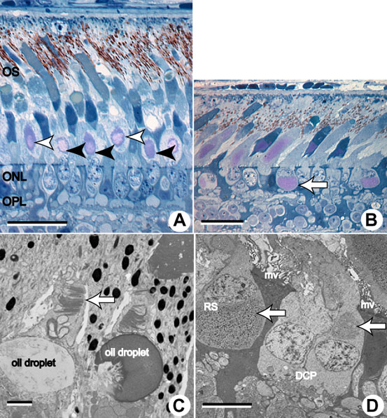
Figure 10. Retinal abnormalities in older birds
Semi-thin sections of retinal samples from rge/rge and control birds at 270 days of age. A: Control retinas show glycogen deposits only in the ISs (external to the outer limiting membrane), which are associated with the rod hyperboloid (white arrowheads) and with the cone accessory cell paraboloid (black arrowheads). The bar represents 20 μm. B: Larger glycogen deposits were observed at this age in retinas from rge/rge birds, that quite often were displaced internal to the outer limiting membrane of the accessory cells of the double cones (arrow). These glycogen deposits are metachromatic in toluidine blue stained semi-thin sections and appear pink in color. The OSs are clearly shorter and disorganized. The bar represents 20 μm. C: Ultra-thin section of a retinal sample from an rge/rge bird demonstrating finer detail of the very short and disorganized cone OSs (arrow). The bar represents 1 μm. D: Ultrastructural detail of glycogen deposits (arrows), present in the supranuclear cytoplasm of an accessory cone cell of the double cone pedicle (DCP) and in the subnuclear cytoplasm of a rod cell spherule (RS) of an rge/rge bird. Long Müller cells microvilli (mv) are abundant between the ISs of the photoreceptor cells of this age group. The bar represents 5 μm. The outer segments (OS), outer nuclear layer (ONL), and outer plexiform layer (OPL) are labeled.