![]() Figure 4 of
Talarico, Mol Vis 2005;
11:169-178.
Figure 4 of
Talarico, Mol Vis 2005;
11:169-178.
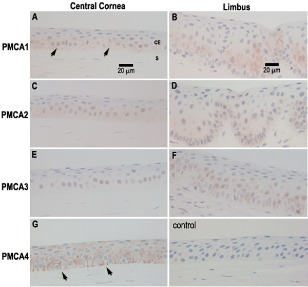
Figure 4. Localization of PMCA Isoforms in paraffin sections of human corneal epithelium from surgical specimens
PMCA1 was found mainly on basal cells in central cornea including the plasma membrane facing the stroma (A; arrows), and in the cytoplasmic domain of basal, and especially wing cells, in the limbus (B). PMCA2 was located mainly on cytoplasmic membranes in basal and wing cells of both central cornea (C) and limbus (D). PMCA3 IR was observed on basal cell nuclei in central cornea (E) and in perinuclear regions of basal and wing cells in the corneoscleral limbus (F). PMCA4 IR was prominent on plasma membranes of cells in all layers of corneal epithelium (G), but appeared to be lacking from most basal cell plasma membranes facing the stroma (G; arrows). Sections reacted with nonimmune rabbit or mouse sera were unstained (the mouse control is shown). Bars in A and B represent 20 μm and apply to all panels. The corneal epithelium (CE) and the stroma (S) are labeled in panel A.