![]() Figure 3 of
Talarico, Mol Vis 2005;
11:169-178.
Figure 3 of
Talarico, Mol Vis 2005;
11:169-178.
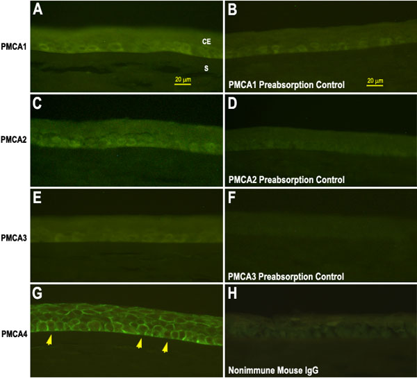
Figure 3. Localization of PMCA isoforms in frozen sections of human corneal epithelium from postmortem specimens
Immunostaining with isoform specific PMCA antibodies showed that PMCA1 (A), PMCA2 (C), and PMCA3 (E) were found primarily in basal cells. PMCA4 IR was localized to plasma membranes in the squamous, wing and basal cell layers, but was lacking along the basal cell membrane adjacent to the stroma (G, arrows). Controls included preabsorption of primary antibodies with the appropriate immunizing peptide for PMCA1 (B), PMCA2 (D), PMCA3 (F), or incubation with nonimmune IgG for JA9 (H). Staining was reduced or eliminated in all control conditions. The bars in (A) and (B) represent 20 μm and apply to all panels. The corneal epithelium (CE) and the stroma (S) are labeled in panel A.