![]() Figure 1 of
Choudhary, Mol Vis 2005;
11:163-168.
Figure 1 of
Choudhary, Mol Vis 2005;
11:163-168.
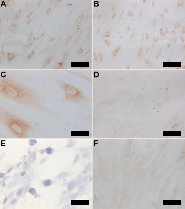
Figure 1. Immunohistochemical staining
A,B: Nonconfluent cultured keratocytes labeled for TSP1 (A) and TSP2 (B) before reaching confluence (DAB staining). C: TSP1 immunoreactivity was localized in a diffuse granular perinuclear pattern with peripheral punctuate staining. D: No staining could be visualized in the control procedures in which primary antibody was replaced by IgG fragments. E: Keratocytes have a rounded configuration at 72 h ai with HSV1 (hematoxylin & eosin). F: A clear reduction in the protein signal (compared to B) is seen for TSP2 4 h ai (DAB staining). The scale bars represent 54 μm in A,B,D,F and 50 μm in C,E.